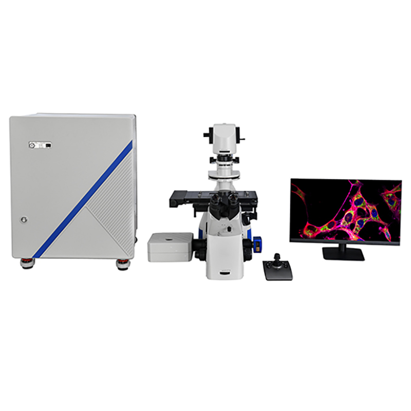
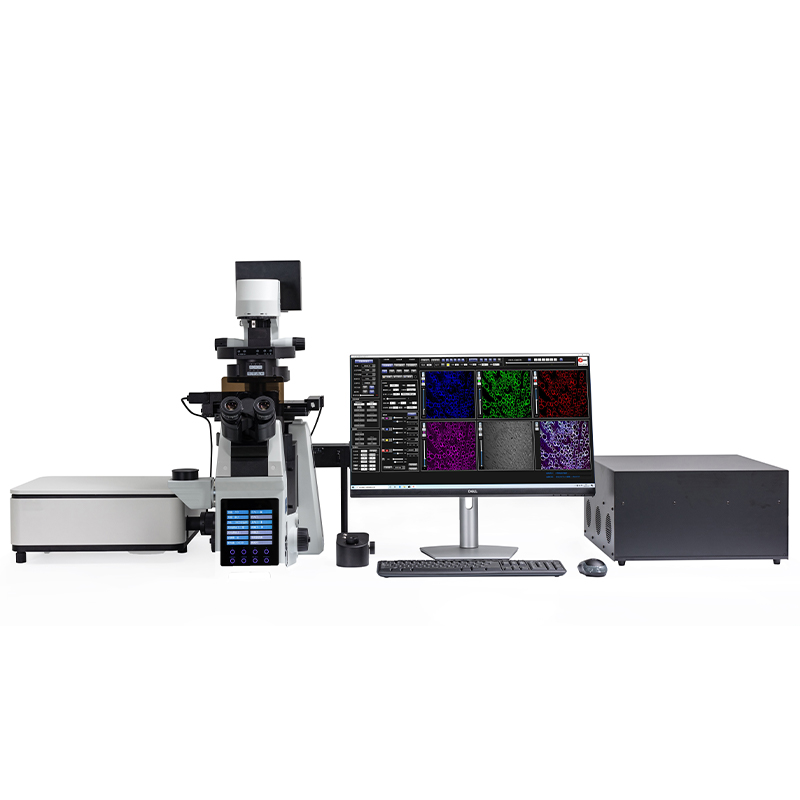


BCF297 Laser Scanning Confocal Microscopy
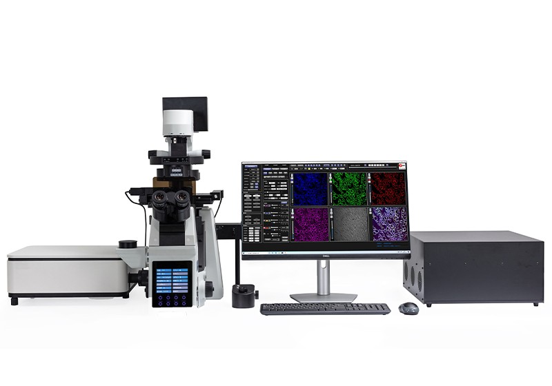
BCF297 is a newly launched laser scanning confocal microscope, which can achieve high-precision observation and precise analysis. It can be widely used in morphology, physiology, immunology, genetics and other fields. It is an ideal partner for cutting-edge biomedical research.
Features
1. High signal-to-noise ratio.
High-efficiency confocal imaging optical path can provide fluorescence images with extremely high signal-to-noise ratio even under weak fluorescence.
2. Excellent image.
Wide spectrum, high numerical aperture lens, perfect for shooting various types of confocal samples.
3. Easy to use.
Full electric frame, optimized design of human-computer interaction interface, allowing you to do a job with ease during sample shooting.
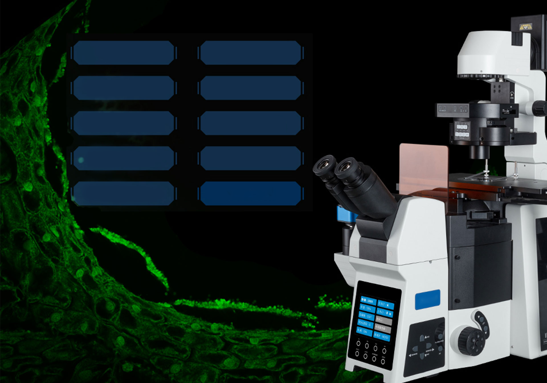
4. All motorized Control System.
The Z-axis of BCF297 laser confocal microscope adopts a motorized device, which can quickly adjust the Z-axis height according to the real-time image. AF One-button autofocus, eliminating the need for fine-tuning steps and improving work efficiency.
The integrated control buttons on both sides of the body can realize the quick switching or rotation of the condenser, brightness, objective lens, attenuation film turntable, and fluorescence turntable, which improves the convenience of operation.
5. Superior Optics for Confocal Imaging.
(1) Unique imaging pinhole structure: minimize the interference caused by component displacement and improve the signal-to-noise ratio and axial resolution of the image.
(2) Controller detection unit: high-sensitivity detection unit (maximum QE≥45%@500nm), which can automatically complete multi-color fluorescence confocal imaging conveniently and quickly.

6. Two sets of optional objective lenses.
BCF297 laser scanning confocal microscope has two sets of optional objective lenses, apochromatic (2X-100X) and super-apochromatic (10X-100X), with a wide range of magnification coverage, suitable for advanced research microscopy and microscopic image shooting. Large numerical aperture further improves resolution.
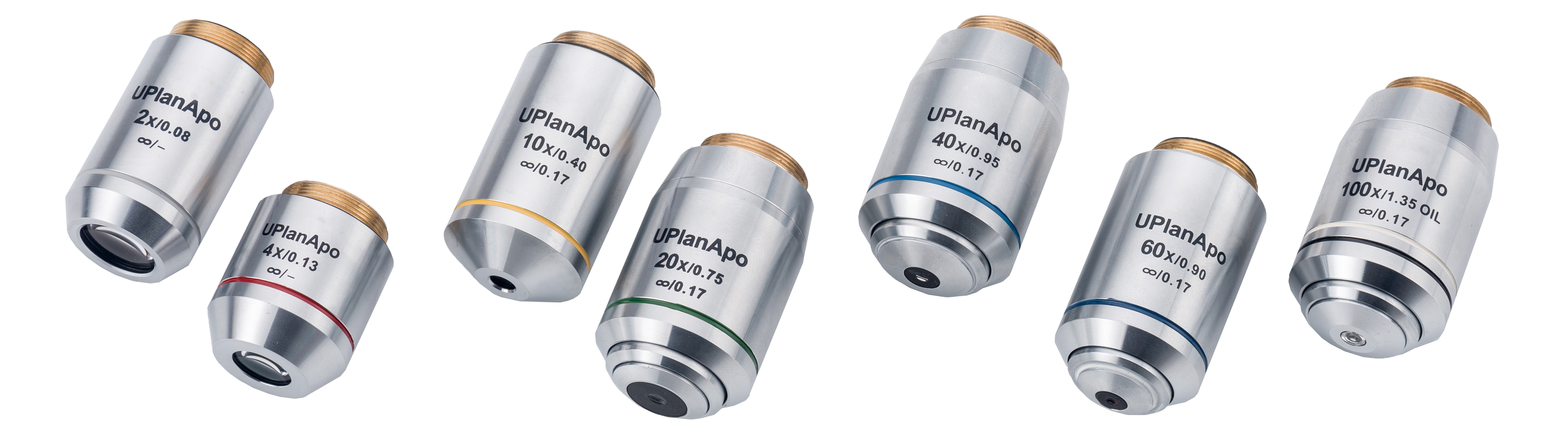
Infinite Plan APO objective
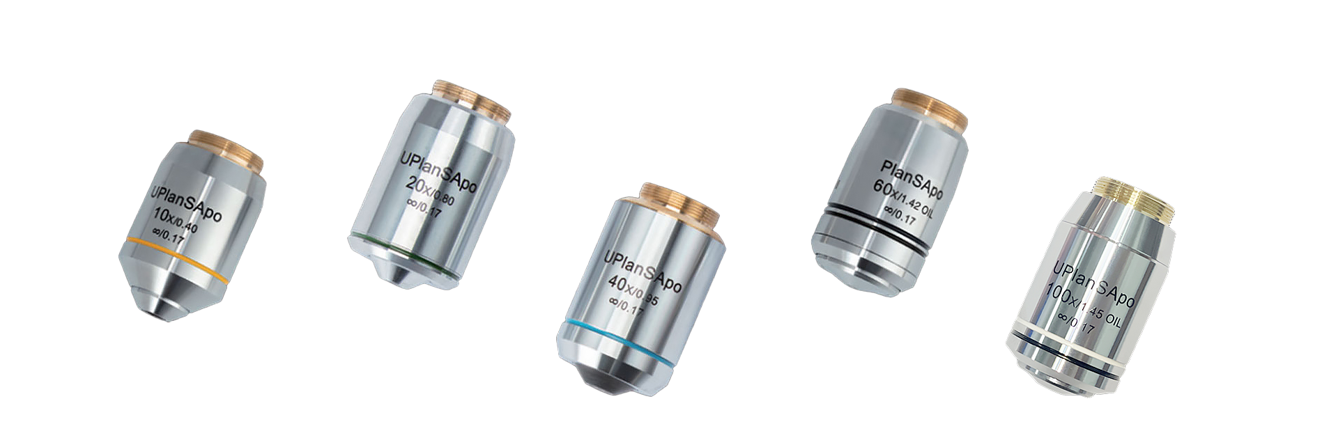
Infinite Plan Super APO objective
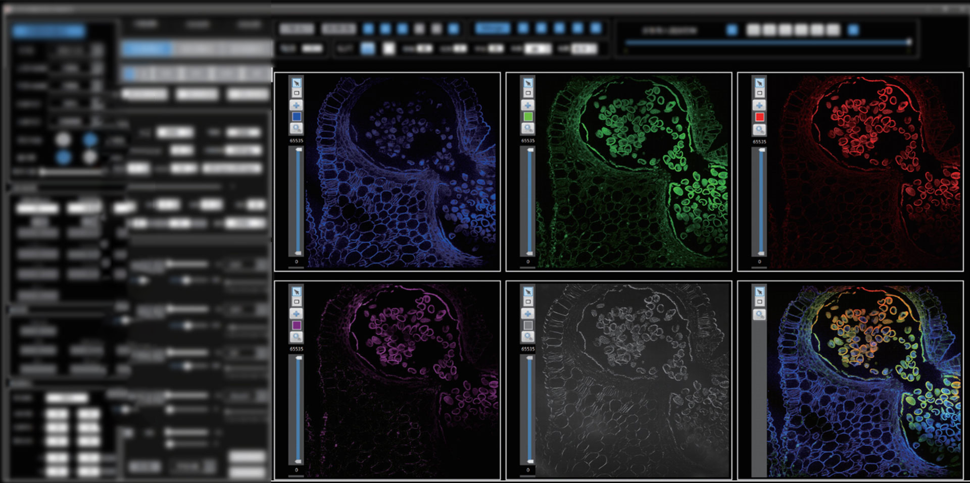
7. Special software for laser confocal.
The software for this confocal microscope is packed with powerful features. BCF297 supports single-channel or multi-channel 2D imaging (XY), 3D imaging (XYZ), 4D imaging (XYZT) and multi-site scanning. Imaging, photobleaching and photostimulation can be performed in user-defined ROI (Region of Interest), Z-Stack imaging, large image stitching, scale correction, filter processing, data recording, etc. can also be performed.
Specification
|
Item |
Specification |
BCF297 |
|
| Laser light source | Laser | 405nm/50mW, 488nm/50mW, 561nm/50mW, 640nm/40mW |
● |
| Laser control | Direct modulation and acousto-optic modulation, all laser switches and intensity can be directly adjusted, and can automatically enter the off state if it is not used for a short time |
● |
|
| Scan module | Scanning unit | Dual-axis XY high-speed optical scanning galvanometer |
● |
| Field of view 14mm×14mm (≥ 19) |
● |
||
| Scanning pixels 512×512 ~ 4096×4096 |
● |
||
| Pixel time 0.5μs ~ 8μs |
● |
||
| Standard scanning speed: 1fps (512×512, 2μs) Fast scanning speed: 3fps (512×512, 0.5μs) |
● |
||
| Zoom scan: 1-32X |
● |
||
| Pinhole | Ф30/40/50μm circular pinhole, located between the dichroic beam splitter and the scanning galvanometer, to ensure that the fluorescence collection efficiency of the system is always maintained at 100% |
● |
|
| Dichroic mirrors and filters | Four-channel dichroic beam splitter: 405/488/561/640nm |
● |
|
| 6-position motorized filter wheel Comes standard with four: 445nm/40, 530nm/43, 607nm/36 and 685nm/40 |
● |
||
| Detection unit | MA PMT, QE ≥ 25%@500nm, 20%@600nm (PMT quantity 1) |
● |
|
| GaAsP PMT, QE ≥ 45%@500nm, 40%@600nm |
○ |
||
| DIC detection unit | With differential interference imaging function, it can realize "DIC-fluorescence" imaging |
● |
|
| Research Grade Inverted Microscope | Optical system | Infinity Chromatic Aberration Corrected Optical System |
● |
| Observation head | 20°-45° adjustable inclination, inverted image, infinity hinged binocular observation tube, interpupillary distance adjustment range: 50~76mm |
● |
|
| Eyepiece | High eye point wide field plan eyepiece PL10X/22mm, adjustable diopter, micrometer |
● |
|
| Objective lens | Infinity Plan Apochromat Objectives 4x, 10x, 20x, 40x, 60x, 100x |
● |
|
| Infinity Plan Super Apochromat Objectives 10x, 20x, 40x, 60x, 100x |
○ |
||
| Microscope frame | Low hand position coarse and micro coaxial electric drive focusing mechanism, Z-axis stroke 10.5mm, precision 1um; front large-size LCD touch display panel, integrated electric control buttons on the body, which can realize condenser, brightness, objective lens, optical port, etc. Quick switch between functions. Built-in motorized glazing port, located on the left side of the fuselage, split ratio 100:0, 0:100; built-in motorized left port, split ratio 0:100, 50:50, 100:0; single-layer/double-layer optical path optional , providing room for system expansion, and one-key switching of optical path modules of each layer according to needs. With Fluorescent Visor, 6-Position Motorized Converter (with DIC Slot) and Glazed Port CTV Adapter Kit |
● |
|
| Stage | Manual mechanical platform, table size 300mm(X)×240mm(Y), moving range 135mm(X)×85mm(Y)
Electric stage, table size not less than 260mm(X)×153mm(Y), moving range 110mm(X)×75mm(Y), with independent operating handle; maximum speed 3mm/S, repeat positioning accuracy ±1um; Equipped with 35mm petri dish, section observation |
○ |
|
| Condenser | Motorized 7-hole condenser, NA 0.55, WD 27mm; 3 holes for φ30mm (phase contrast), 4 holes for φ38mm (DIC); support bright field, phase contrast, DIC observation (including polarizer set) |
● |
|
| Fluorescent lighting system | 8-hole fluorescent turntable system, the system can determine the position of the turntable placed on the upper and lower layers of the rack; it also has an electric shutter function, which can directly block the fluorescent lighting source, with fluorescent filter block mirror group: B/G/UV, etc. |
● |
|
| Operating software
|
Scanning imaging | Image acquisition and system automatic control function, full electric control switching of optical path; self-adaptation of camera parameters and preview parameters; full field of view and ROI scanning imaging; single-color or multi-color 2D imaging (XY), 3D imaging (XYZ), 4D imaging (XYZT) and multi-site scanning |
● |
| Processing analysis | Multi-color fluorescence colocalization processing, fluorescence and differential interference interference (DIC) image superposition; calibration and adding scales; filtering processing; Z-Stack processing analysis and large image stitching |
● |
|
| Data management | A variety of hardware connections are optional, automatic image storage paths can be set, multiple image output formats, and scanning imaging parameters can be automatically saved |
● |
|
Note: ●Standard Outfit, ○Optional
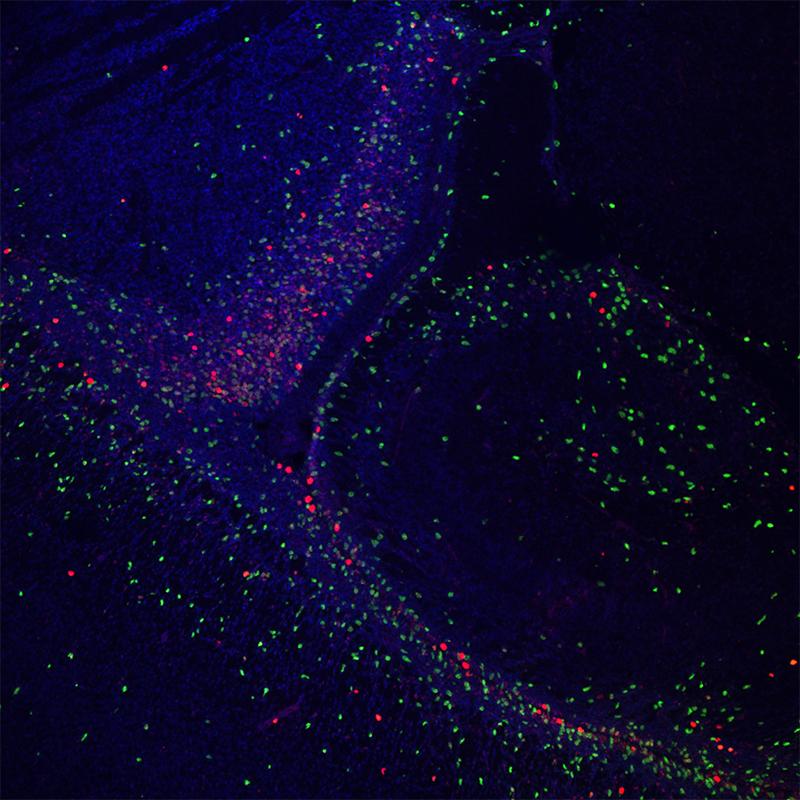
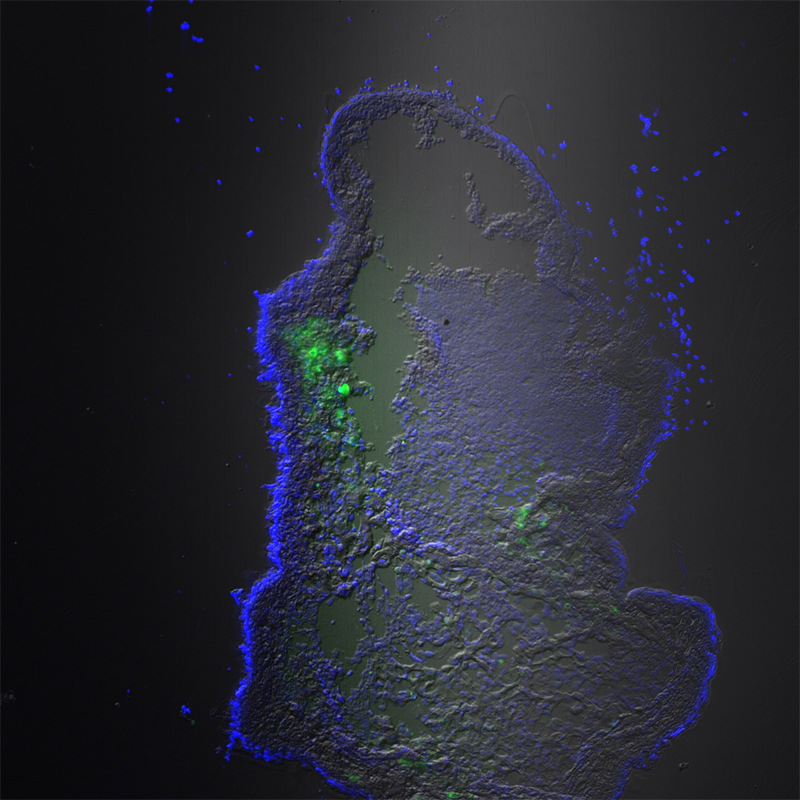
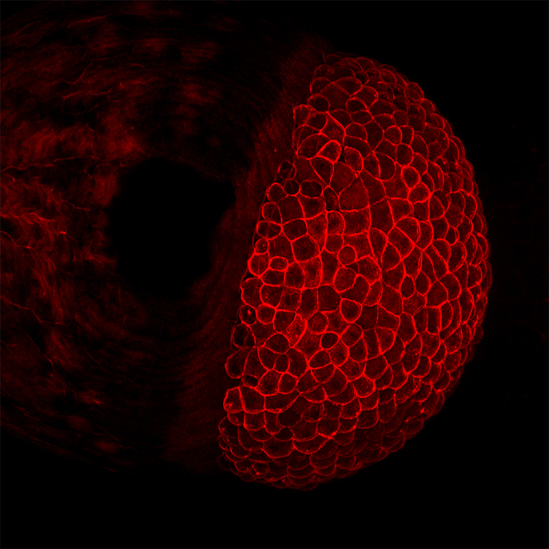
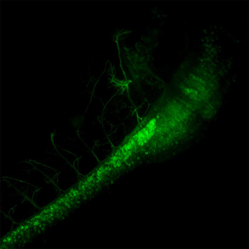
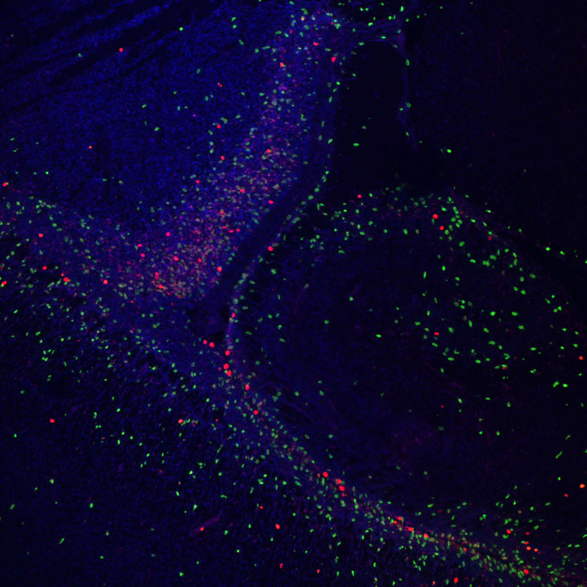
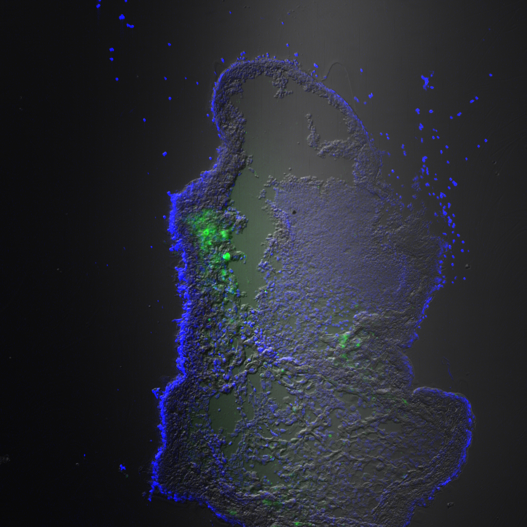
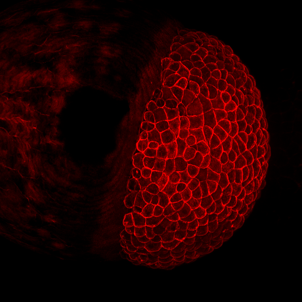
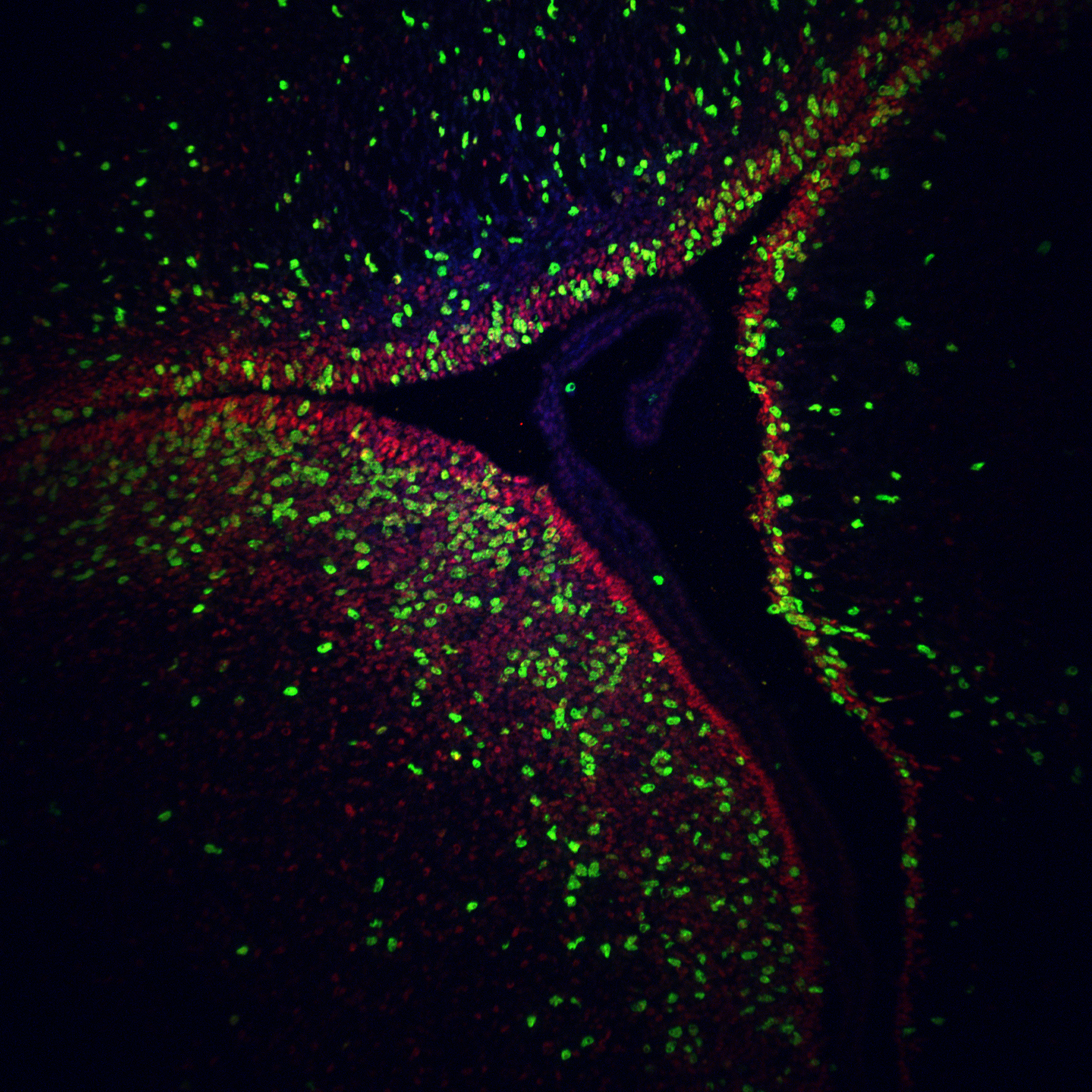
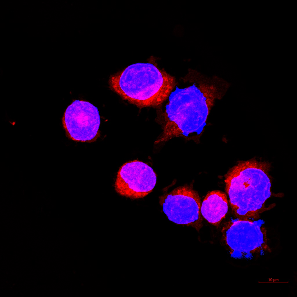
Sample Images

Certificate

Logistics