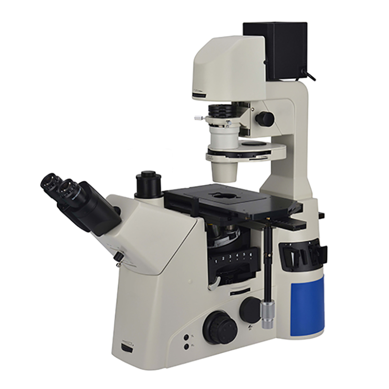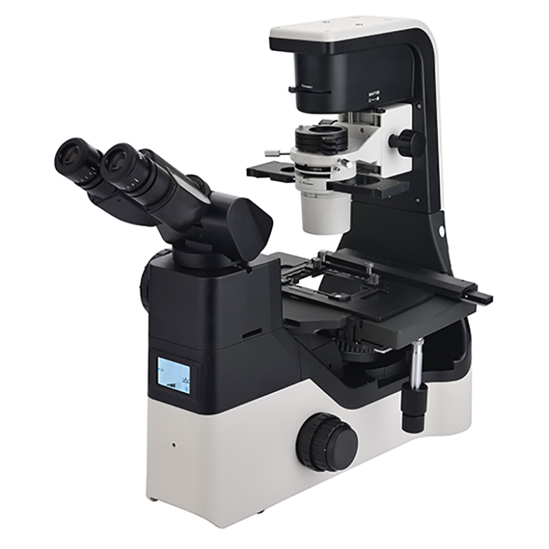


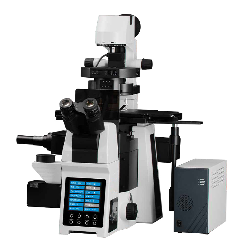
B-SIM298 Structured Illumination Fluorescence Microscope
B-SIM298
Introduction
B-SIM298 Fluorescence microscope integrates observation and analysis. Automated control of imaging process, excellent for live cell imaging. B-SIM298 has powerful expansion functions, providing a more complete solution for life science research.
Feature
1.High optical resolution
X/Y axis resolution ratio: 240nm; Z-axis resolution ratio: 600nm.
2.High sensitivity backlit sCMOS camera
High sensitivity backlit sCMOS camera, with a frame rate of up to 100 frames per second and a quantum efficiency of up to 95% at 2048x2048 pixels, with a 6.5um pixel size.
3.Illumination
4-wavelength LED light source, long life, high brightness, low light toxicity, high uniformity lighting solutions.
4.Software
Hardware control: 12-axis fully electric control, camera control, supports third-party light source, electric stage and other peripheral control.
Image acquisition: x, y, z, λ, t, n, l, seven-dimensional acquisition, multi-dimensional, full process automated control.
Image processing: 3D reconstruction and display, co-localization processing, co-localization linkage, image brightness, contrast, threshold processing, image flipping, mirroring, background removal, dynamic image generation, stack processing and ROI processing.
Image analysis: distance, perimeter, area, roundness, maximum and minimum gray scale, and other parameter analysis, co-localization analysis, cell counting, particle counting, protein tracking, subpopulation analysis and cell cycle analysis.
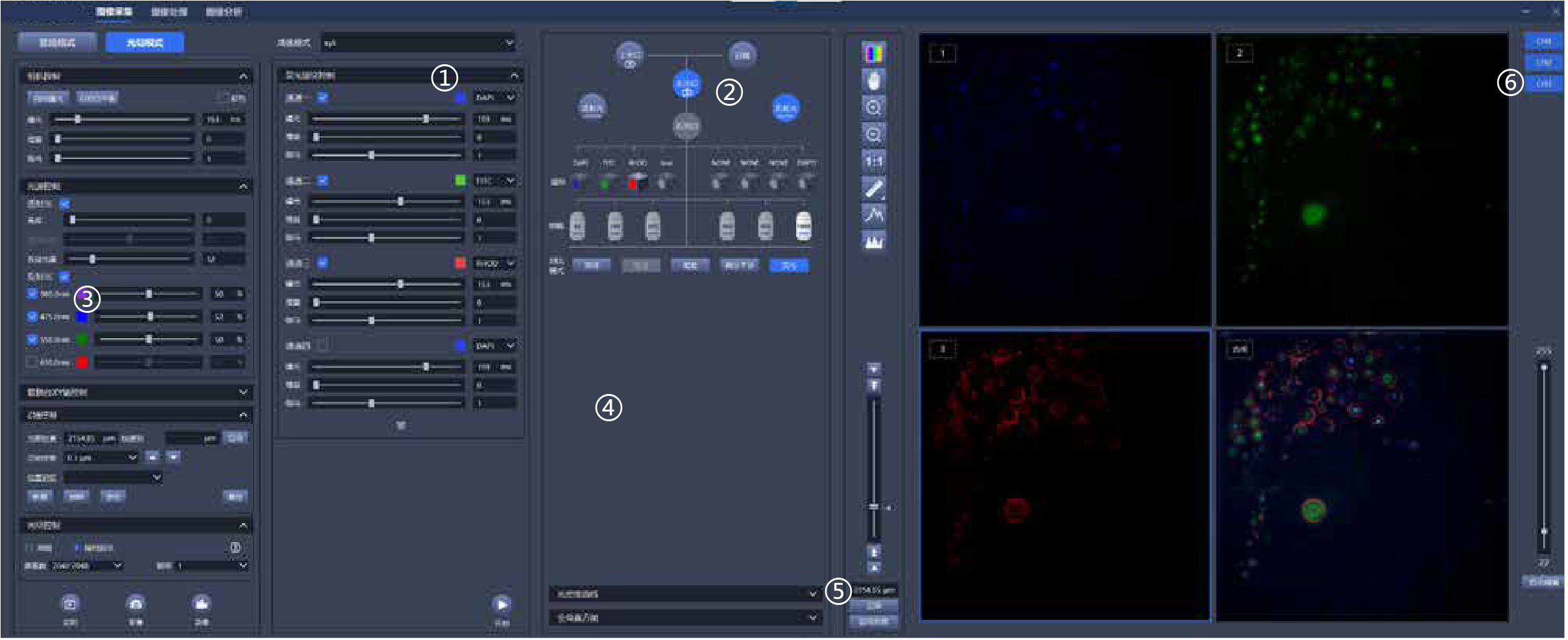
1.Five-dimensional sequence acquisition of x, y, z, λ, t, realizes automatic acquisition control.
2.Graphical interaction: switch optical port, filter block, objectives with one-click and changing observation mode.
3.Electric focusing, electric stage, electric fluorescence filter block, electric condenser, electric light source control, electric light path switching, realizing fully electric 12-axis control.
4.Real-time optical density measurement, real-time global histogram display.
5.Graphical focus control, focus position memory, automatic focus control.
6.Customize 1-8 channel acquisition, real-time pseudo-color display, arbitrary multi-channel overlay, and real-time display of multi-channel overlay map.
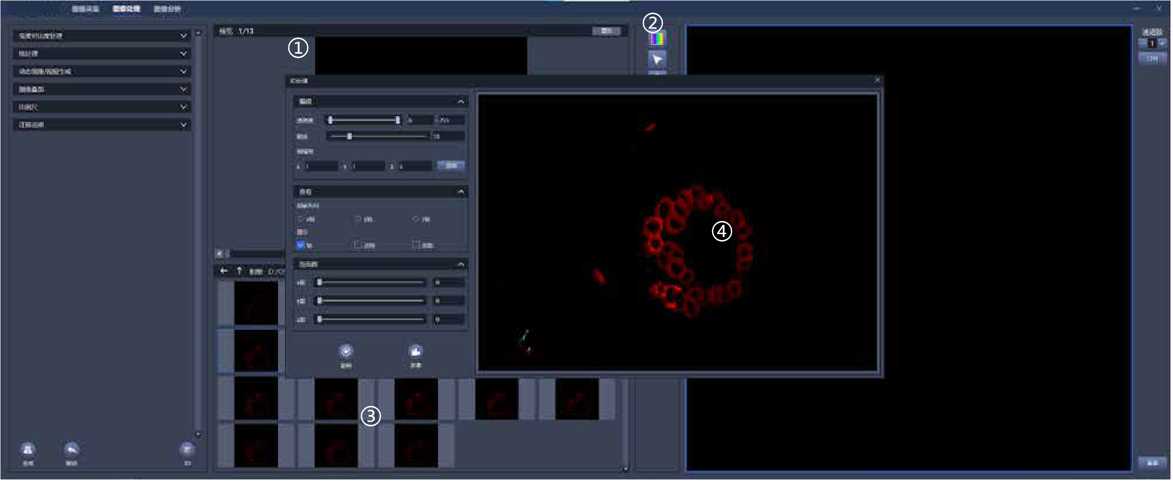
1.Real-time display of comparison before and after image processing
2.Record image information in detail, including detailed parameters such as acquisition channel, objectives, exposure time, etc.
3.Real-time image preview
4.3D image reconstruction
Specification
| Item | Specification | B-SIM298 | |
| Optical System | Infinity Color Correction Optical System | ● | |
| Viewing Head | High eye point wide field plan eyepiece PL10X/22mm, with adjustable diopter | ● | |
| Eyepiece | 20-45 degree tilting binocular tube, interpupillary distance: 50-76mm | ● | |
| Objectives | Long Working Distance Plan Semi- Apochromatic Objectives | 4X/NA=0.13, WD=17mm | ○ |
| 10X/NA=0.3, WD=8.8mm | ○ | ||
| 20X/NA=0.45, WD=6.5-7.6mm, coverslip thickness: 0-2mm | ● | ||
| 40X/NA=0.6, WD=2.85-4.05mm, coverslip thickness: 0-2mm | ● | ||
| 60X/NA=0.7, WD=1.42-2.1mm, coverslip thickness: 0-1.3mm | ○ | ||
| Infinity Plan Semi-Apochromatic Objectives | 4X/NA=0.16, WD=12.8mm | ● | |
| Infinity Plan Super Apochromatic Objectives | 10X/NA=0.4, WD=3.1mm | ● | |
| 20X/NA=0.8, WD=0.6mm | ○ | ||
| 40X/NA=0.95, WD=0.18mm | ○ | ||
| (Oil) 60X/NA=1.42, WD=0.17mm | ● | ||
| (Oil) 100X/NA=1.45, WD=0.13mm | ○ | ||
| Microscope Body & Nosepiece | Low position coarse and fine coaxial electric focusing mechanism, range: 10.5mm, precision: 1μm. Built-in electric upper camera port, splitting ratio: 100:0 / 0:100. Built-in electric left camera port, splitting ratio: 0:100 / 50:50 / 100:0, dual optical path, with fluorescent light barrier. Electric bright field sextuple nosepiece with DIC slot and upper optical port CTV adapter. | ● | |
| Base mounting bracket | ○ | ||
| Right camera port, splitting ratio: 100:0 / 0:100, field of view: 16mm. Built-in 1X CTV, C-mount adapter | ○ | ||
| Illumination | Pillar tilt mechanism, Koehler transmission illuminator, adjustable condenser holder with 65mm stroke. 4 filters holders with LBD, Green filter, Neutral filter for halogen models or Neutral filter for LED models | ● | |
| 12V/100W halogen illumination, filament center preset | ○ | ||
| 12V/100W halogen lamp | ○ | ||
| 10W cool color LED light illumination, color temperature 5000K | ● | ||
| Condenser & Iris | Electric septuple condenser, NA 0.55, WD=27mm. 3 holds for Φ30mm (phase contrast), 4 holds for Φ38mm (DIC), support for bright field/phase contrast /DIC (with polarizing kit) | ● | |
| Super long working distance manual condenser with 5 holes, NA 0.3, WD=73mm, support for 4X-60X phase contrast, simple polarizing observation and 10X-40X relief phase contrast observation. | ○ | ||
| Cable 100cm | ● | ||
| DIC | Transmitted DIC kit | ● | |
| 10X transmitted DIC ring (for BSIM2980022) | ● | ||
| 20X transmitted DIC ring (for BSIM2980022) | ● | ||
| 40X/60X transmitted DIC ring (for BSIM2980022) | ● | ||
| Analyzer kit (for BSIM2980022) | ● | ||
| Fluorescent Module | Fluorescence attachment with 8 holes, with electric shutter | ● | |
| Cable 30cm, connecting the fluorescence attachment to the frame | ● | ||
| Dust cap | ● | ||
| UV fluorescence filter house, EX: AT375/28X, 25mm, BS:AT415DC, 25.5*36*1mm, EM:AT460/50M, 25mm | ● | ||
| B fluorescence filter house, EX: AT480/30X, 25mm, BS: AT505DC, 25.5*36*1mm, EM: AT535/40M, 25mm | ● | ||
| G fluorescence filter house, EX: AT560/40X, 25mm, BS: AT600DC, 25.5*36*1mm, EM: AT635/60M, 25mm | ● | ||
| G fluorescence filter house, EX: AT620/50X, 25mm, BS: AT655DC, 25.5*36*1mm, EM: AT690/50M,25mm | ● | ||
| LED light source: 380/475/550/630 four wavelengths, four independent lamp beads, high-power, long-life LED light source. | ● | ||
| Stage | Manual mechanical stage, size: 300mm(X)*240mm(Y), moving range: 135mm(X)*85mm(Y), stage thickness: 30mm. Right universal handle, X/Y axis limitable and lockable, moving range 50mm * 50mm after locked; with pressure clap for holding slices and culture flasks, with Φ110mm replaceable disc (inner Φ30), with metal stage plate with waist shaped hole. | ● | |
| Electric Control Box & PC | Electric control box, input voltage 90-265VAC wide voltage, output 12V100W or 12V10W.Digitally adjustable output voltage through CAN, in addition to three outputs of 24V5A/15V5A/5V5A, equipped with forced air cooling, including one 3C power cord. | ● | |
| DB26 Cable 200cm, connecting the electric control box to the frame | ● | ||
| DELL 3020T computer: I7-12700 32GB RAM 1TB solid state, host RTX3060TI+8G independent display, Dell 34 display, curved USB-C display iS3423DWC, WIN10 | ● | ||
| DB9 Cable 200cm, connecting the PC to the electric control box | ● | ||
| USB-CAN card. When the customer purchases computer by themselves and requires computer to control microscope electric control, it must be paired. | ● | ||
| Structured Light Components | Grid size: 40um. Light transmission range: 400-750nm; liquid core fiber: core diameter: 3mm. Numerical aperture: 0.5. Transmission range: 240-740nm | ● | |
| Camera | sCMOS back-illuminated black and white camera. Global pixels: 2048*2048, 4 million effective physical pixels. Pixel size: 6.5μm*6.5μm. Chip size: 13.3mm*13.3mm. Quantum efficiency: 95% @ 600nm. Frame rate: 100fps@CameraLink, 40fps@USB 3.0. Full well capacity: typical: 45 ke-. Dynamic range: 90dB. Readout noise: CMS: 1.1E-Median, 1.2E-RMS. Exposure time: 6.6μs-10s.Refrigeration method: air cooling, water cooling. Dark current: air cooling: 0.15e-/pixel/s@-15℃, water cooling: 0.10e-/pixel/s@-25℃. Bit depth: 11bit, 12bit, 16bit. | ● | |
| Hardware Control | Hardware control: Automatically control the structured light lighting system. Image acquisition: exposure time control, gain control, threshold control, gamma value control, image pixel number control, x, y, z, λ, t five-dimensional acquisition, custom xyz, xyt, xyzt, xyλ, xyλt, xyzλt, multiple collection modes. Start recording with one click. Supports real-time pseudo-color annotation and real-time optical density measurement. Image processing: 3D reconstruction and display, image flipping, mirroring, background removal, dynamic image generation, stack processing, ROI processing. Image analysis: perimeter, area, roundness, maximum gray level, minimum gray level and other parameter analysis, co-localization analysis, cell counting, point counting, protein tracking, subpopulation analysis. |
● | |
| Telescope | Telescope (Φ30) | ○ | |
| Adapter | 1X C-mount adapter, adjustable focus | ● | |
| Other Accessories | Fluorescent free oil 30ml | ● | |
| Internal hexagonal Spanner M3 for phase contrast adjusting screw | ● | ||
| Internal hexagonal Spanner M4 | ● | ||
| Internal hexagonal Spanner M5 | ● | ||
Note: ● Standard Outfit, ○ Optional
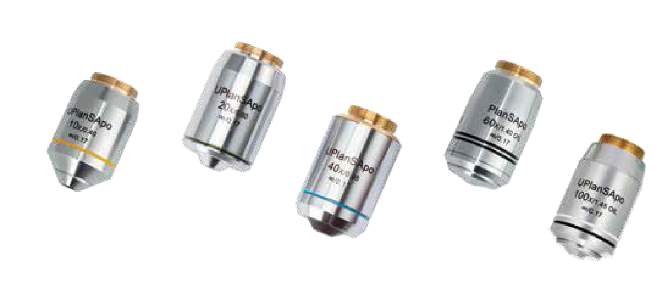
Accessories
Infinity plan super apochromatic objectives
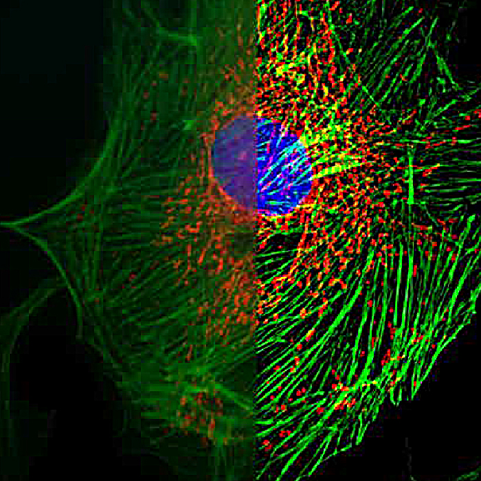
Sample Images
Certificate
Logistics